Shimadzu iMScope AP-MALDI QTOF
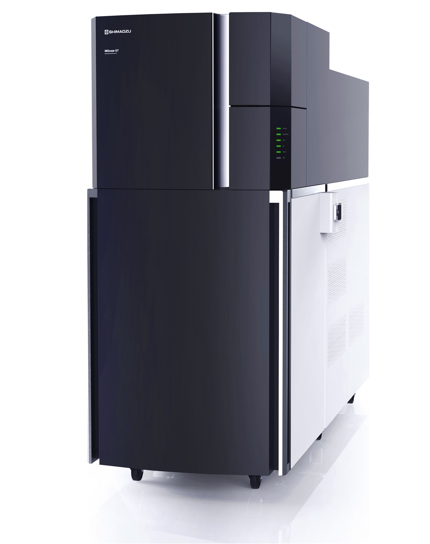 The Shimadzu iMScope AP-MALDI QTOF is specialized for imaging mass spectrometry (IMS) applications where information on the spatial distributions of analytes is required. The iMScope has an atmospheric pressure MALDI source which eliminates vacuum equilibration making sample intnroduction fast. The built-in optical microscope (5X, 10X, 40X) allows for direct visualization of the histology sections and easy selection of ROIs for imaging. The 20kHz Nd:YAG laser acquires data at speeds up to 50 pixels/sec at a resolution as low as 5 µm although 10-20 µm is more practical. Mouse brain images (1 cm2) can be acquired within 1-4 hours. Accessory equipment available in the facility includes a Leica CM1850 cryostat for cryosectioning onto indium titanium oxide (ITO) conductive slides. Matrix application can be applied with the HTX M3+ matrix sprayer or Shimadzu iMayer sublimator. Users can select from standard pre-programmed methods. MS data are processed with Shimadzu's ImageReveal or Bruker's SCiLS Lab Pro software. Remote access for data processing is available.
Access: staff operated and validated users
Analyzer: AP-MALDI QTOF
Mode: Positive or negative
Sample prep: histology sections or other thin flat specimens on ITO slides or metal target plates
Sample plate: 2 standard size (3"x1") ITO slides or Shimadzu sample holder
Software: ImageReveal (Shimadzu) and SCiLS Lab Pro (Bruker)
Resources/Protocols
Recent Publications:
- Spatially resolved detection of small molecules from press-dried plant tissue using MALDI imaging. Zane G. Long, Jonathan V. Le, Benjamin B. Katz, Belen G. Lopez, Emily D. Tenenbaum, Bonnie Semmling, Ryan J. Schmidt, Felix Grün, Carter T. Butts, and Rachel W. Martin, Applications in Plant Sciences in press (2023).
Poster abstract: https://doi.org/10.1016/j.bpj.2022.11.1560
Bruker ultrafleXtreme MALDI-TOF
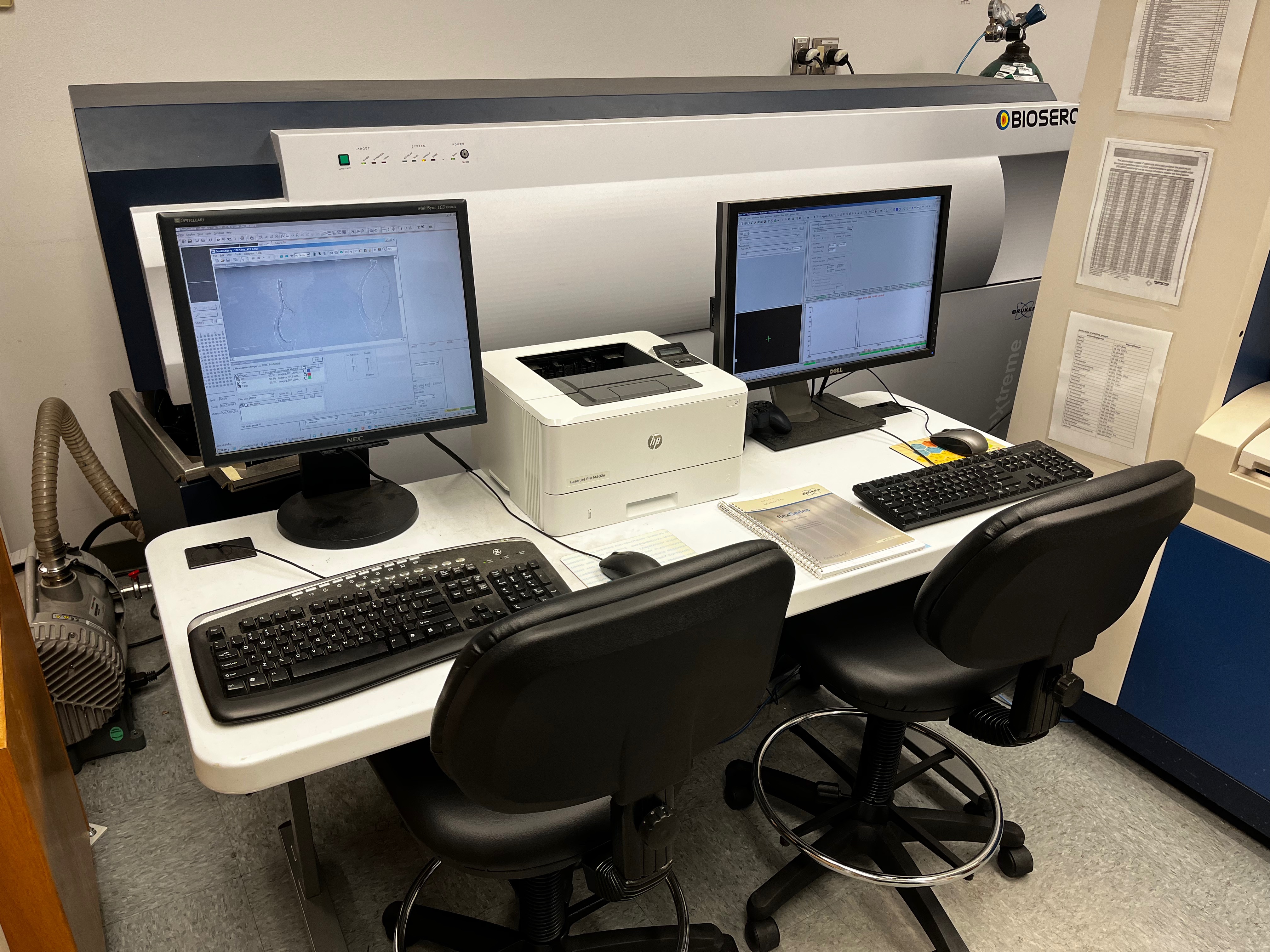
The Bruker ultrafleXtreme MALDI-TOF is principally dedicated for MALDI imaging mass spectrometry (IMS) applications where information on the spatial distributions of analytes is required. Biological histology samples are cryosectioned (10-20 µm thickness) onto indium titanium oxide (ITO) slides, coated with matrix compounds by spray deposition or sublimation, and MALDI data acquired by laser spot rasterization (spacing step size 10-100 µm). Imaging software (flexImaging, SCiLS Lab Pro or ImageReveal) generates images colored for relative analyte concentrations and performs statistical analyses (ROIs, PCA, cross-correlation, segmentation, classification etc.).
Access: staff operated and validated users
Analyzer: MALDI TOF
Mode: Positive or negative
Sample prep: histology sections or other thin flat specimens on ITO slides or metal target plates
Sample plate: 2 standard size (3"x1") ITO slides or Bruker sample plate
Software: flexControl, flexImaging, flexAnalysis, SCiLS Lab Pro (Bruker), ImageReveal (Shimadzu)
Resources/Protocols
Recent Publications:
- Spatially resolved detection of small molecules from press-dried plant tissue using MALDI imaging. Zane G. Long, Jonathan V. Le, Benjamin B. Katz, Belen G. Lopez, Emily D. Tenenbaum, Bonnie Semmling, Ryan J. Schmidt, Felix Grün, Carter T. Butts, and Rachel W. Martin, Applications in Plant Sciences in press (2023).
Poster abstract: https://doi.org/10.1016/j.bpj.2022.11.1560
AB Sciex 5800 MALDI TOF/TOF
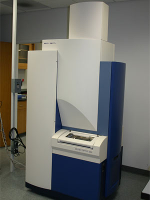
The ABSciex MALDI-TOF/TOF is used for matrix assisted laser desorption ionization (MALDI) analysis of large molecular weight polymers (peptides, nucleic acids, polymeric species).
Peptide/protein/polymer samples are spotted with matrix (DHC, CHCA, DAN, THAP etc.) onto target plates and dried.
Access: Walk-up Open Access
Access: Walk-up Open Access
Analyzer: TOF-TOF
Mode: Positive or negative
Sample prep: Matrix/sample deposition on metal target plates
Sample plate: 100 samples/plate
Software: Voyager software